Laser ultrasound method achieves non-contact medical imaging
Because the new technique does not rely on the use of an ultrasound probe, the read-out technique is not subject to image variability (upon probe pressure and orientation), a major challenge in modern ultrasound imaging.
Here the ultrasound waves are generated remotely by a pulsed laser light tuned at a particular wavelength to penetrates the skin and to be absorbed by blood vessels. The blood vessels rapidly expand and relax, instantly heated by a laser pulse then rapidly cooled by the body back to their original size, only to be struck again by another light pulse. The resulting mechanical vibrations generate sound waves that travel through the skin, bouncing off muscle, fat, and other soft tissues before reflecting back to the skin.
The researchers used a second laser to remotely detect the reflected waves (through the Doppler effect) and translate them into an image similar to conventional ultrasound. This contactless ultrasound imaging technique may help remotely image and assess health of infants, burn victims, and accident survivors in hard-to-reach places. Scanning the forearms of several volunteers, the authors of the paper report the observation of common tissue features such as muscle, fat, and bone, down to about 6 centimetres below the skin, all done from half a meter away.
Although at this point, the laser ultrasound (LUS) images are only comparable to images achieved at the early stages of medical ultrasound imaging decades ago, the researchers are confident those could be drastically improved by leveraging known beam forming and image processing techniques already used for ultrasound imaging.

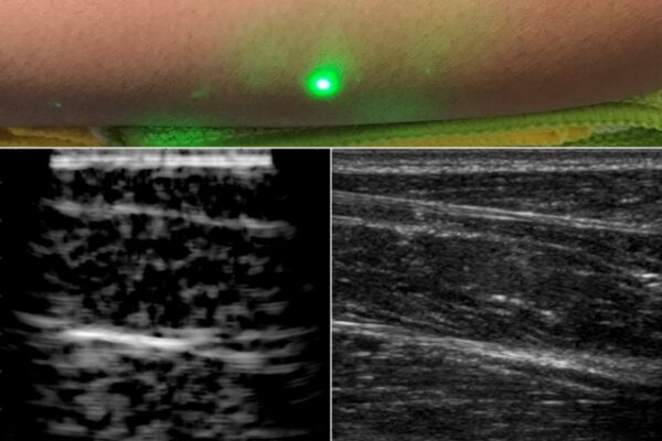
beneath the skin, without making contact with the skin as
conventional ultrasound probes do. The new laser ultrasound
technique was used to produce an image (left) of a human
forearm (above), which was also imaged using conventional
ultrasound (right). Image courtesy of the researchers.
Beyond the use of a fast amplitude-modulated optical source to improve the LUS imaging depth, bandwidth and resolution, the parallelization of optical sources and receivers could enable optical transmit and receive beam forming techniques. This combined with modern computer vision techniques such as 3D imaging and tracking could make large-volume LUS imaging and optical beam forming feasible, note the authors.
With optical spot tracking and a sufficiently fast data acquisition and coverage, LUS systems could enable the simultaneous 3D imaging of both the external and internal tissue geometries, the paper concludes. Indeed, a lot of the technologies required for such 3D acquisitions has already been developed for lidars, with chip-scale steerable laser technology used in autonomous cars.
MIT – www.mit.edu
 If you enjoyed this article, you will like the following ones: don't miss them by subscribing to :